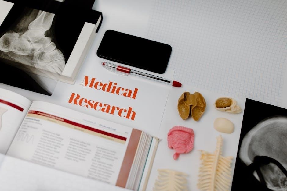
This laboratory manual provides a comprehensive guide for exploring human anatomy and physiology through hands-on activities and practical experiments, fostering deeper understanding and application of theoretical knowledge.
1.1. Overview of the Laboratory Manual
This laboratory manual is designed to complement anatomy and physiology coursework, offering structured exercises and investigations. It provides clear instructions and visual aids to guide students through hands-on activities, fostering practical understanding. The manual is divided into sections, each focusing on specific body systems, such as skeletal, muscular, and nervous systems. It includes dissections, histological studies, and physiological measurements, ensuring a comprehensive learning experience. By integrating theoretical knowledge with practical application, the manual equips students with essential skills for analyzing and understanding human anatomy and physiology effectively.
1.2. Importance of Laboratory Work in Anatomy & Physiology
Laboratory work is essential for understanding anatomy and physiology, as it provides hands-on experience with biological structures and processes. Through dissections, histological examinations, and physiological experiments, students gain practical skills and a deeper appreciation of human biology. Lab activities bridge the gap between theoretical knowledge and real-world applications, fostering critical thinking and problem-solving abilities. By engaging directly with specimens and data, students develop a more comprehensive understanding of bodily functions and systems, preparing them for careers in healthcare, research, and related fields where practical expertise is crucial.
1.3. Safety Guidelines and Precautions
Adhering to safety guidelines is crucial in anatomy and physiology labs to prevent accidents and ensure a safe learning environment. Students must wear protective gear such as gloves, goggles, and lab coats when handling biological specimens or chemicals. Proper handling and disposal of materials are essential to avoid exposure to pathogens or hazardous substances. Emergency procedures, such as knowing the location of fire extinguishers and first aid kits, should be understood by all participants. Following established protocols and instructor instructions is vital to maintaining a secure and efficient laboratory setting.
Essential Laboratory Tools and Equipment
This section introduces the fundamental tools and equipment required for anatomy and physiology labs, including microscopes, dissection tools, measurement devices, and protective gear like gloves and goggles.
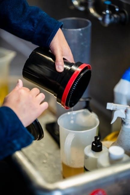
2.1. Microscopes and Histological Tools
Microscopes are indispensable in anatomy and physiology labs for examining cellular structures and tissues. Common types include compound microscopes for detailed tissue analysis and stereo microscopes for three-dimensional viewing of specimens. Histological tools such as scalpels, forceps, and slide preparation equipment are essential for preparing tissue samples. Proper handling and maintenance of these tools ensure accurate observations and safety. These instruments are crucial for understanding microscopic structures, enabling students to connect theoretical knowledge with practical insights. Their effective use is vital for successful laboratory investigations in human anatomy and physiology studies.
2.2. Measurement and Data Collection Tools
Measurement tools like thermometers, sphygmomanometers, and EKG machines are essential for assessing physiological parameters such as temperature, blood pressure, and heart activity. Data collection tools, including lab notebooks and digital software, enable precise recording and analysis of experimental results. These tools facilitate accurate measurements, ensuring reliable data for scientific investigation. Proper use of these instruments allows students to gather and interpret data effectively, enhancing their understanding of anatomical and physiological processes. They are fundamental for conducting experiments and drawing meaningful conclusions in human anatomy and physiology studies.
2.3. Dissection Tools and Supplies
Dissection tools are essential for anatomical exploration, including scalpels, forceps, scissors, and dissecting needles. These instruments allow precise tissue dissection and examination. Supplies like gloves, dissecting trays, and preservatives ensure safe handling and specimen preparation. Proper use of these tools is critical for effective learning and maintaining laboratory safety. They enable students to explore complex anatomical structures, fostering a deeper understanding of human physiology through hands-on experience. Each tool serves a specific purpose, making them indispensable in anatomy and physiology laboratories.
Anatomical Directions and Body Planes
Anatomical directions and body planes provide a standardized framework for understanding spatial relationships in the human body, aiding in precise dissection and visualization of structures in laboratory settings.
3.1. Understanding Anatomical Terminology
Understanding anatomical terminology is fundamental for effective communication in anatomy and physiology. It involves learning standardized terms that describe body structures, their locations, and spatial relationships. Accurate use of terms like proximal, distal, anterior, and posterior ensures clear descriptions and minimizes confusion. Grasping this language is essential for interpreting scientific texts, performing dissections, and visualizing complex structures. In a laboratory setting, precise terminology aids in identifying organs, tissues, and systems, fostering a deeper appreciation of human anatomy. This foundational knowledge is crucial for both theoretical understanding and practical applications in the lab.
3.2. Exploring Body Planes and Sections
Body planes and sections are essential tools for understanding spatial relationships in anatomy. The three main planes—frontal (coronal), sagittal, and transverse (horizontal)—divide the body into sections for detailed study. Frontal sections run vertically from head to toe, sagittal sections divide the body into left and right, and transverse sections are horizontal slices. These planes help in creating two-dimensional representations of three-dimensional structures, aiding in visualization and identification during dissections and imaging. Mastering body planes is crucial for accurate anatomical descriptions and practical applications in laboratory exercises and medical imaging interpretation.
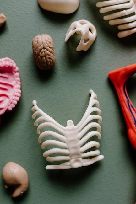
Skeletal System Laboratory Exercises
Explore the skeletal system through hands-on activities, including bone identification, joint analysis, and movement observation to understand bone structure, joint types, and their functional roles in movement.
4.1. Bone Structure and Identification
This section focuses on understanding the structural components of bones, including the periosteum, cortex, spongy bone, and marrow cavity. Students learn to identify bone types—long, short, flat, irregular, and sesamoid—based on shape and function. Hands-on activities involve labeling bone features and analyzing their roles in movement and support. Practical exercises emphasize distinguishing between compact and cancellous bone, observing histological slides, and correlating bone anatomy with its physiological functions. This foundational knowledge is crucial for comprehending skeletal system dynamics and its role in human anatomy and physiology.
4.2. Joint and Movement Analysis
This section explores the classification and functionality of joints, focusing on synovial, cartilaginous, and fibrous types. Students analyze joint movements, including flexion, extension, rotation, and circumduction, using prosected specimens and models. Practical exercises involve palpating joints to identify ligaments and tendons, assessing range of motion, and understanding joint stability versus mobility. The role of joints in facilitating movement while maintaining structural integrity is emphasized, preparing students to apply this knowledge in clinical and biomechanical contexts, such as injury rehabilitation and understanding joint-related pathologies.

Muscular System Laboratory Exercises
This section focuses on identifying and understanding muscle types, their structure, and functions through dissection and palpation. Practical exercises emphasize muscle roles in movement and support.
5.1. Muscle Types and Functions
This section explores the three primary types of muscles: skeletal, smooth, and cardiac. Skeletal muscles enable voluntary movement by contracting bones, while smooth muscles handle involuntary actions like digestion. Cardiac muscles are specialized for pumping blood. Through hands-on dissection and palpation, students identify muscle structures and learn how they contribute to movement and bodily functions. Practical exercises highlight the roles of muscles in maintaining posture, facilitating locomotion, and supporting internal processes. Understanding muscle types and functions is crucial for analyzing human physiology and diagnosing muscle-related disorders.
5.2. Muscle Dissection and Identification
This exercise involves hands-on dissection to identify and examine muscle structures, emphasizing their arrangement, attachments, and fiber composition. Students use dissecting microscopes and probes to explore muscle tissue samples, observing features like tendons, fascicles, and striations. The process includes identifying muscle origins and insertions, understanding their functional significance. Practical identification of skeletal, smooth, and cardiac muscles enhances familiarity with their distinct characteristics. This laboratory activity bridges theoretical knowledge with tactile learning, reinforcing the importance of muscles in movement and bodily functions.
Nervous System Laboratory Exercises
This section explores the structure and function of the nervous system through hands-on activities, including brain and spinal cord dissection, nerve tissue examination, and reflex testing.
6.1. Brain and Spinal Cord Structure
In this section, students examine the brain and spinal cord, focusing on their structural and functional components. Hands-on activities include dissecting brain regions, such as the cerebrum, cerebellum, and brainstem, to identify key features like sulci, gyri, and ventricles. The spinal cord’s anatomy is explored, highlighting the gray and white matter, as well as nerve tracts. Microscopic examinations of nervous tissue reveal neurons and glial cells. These exercises provide a foundational understanding of the central nervous system’s organization and its role in controlling bodily functions, emphasizing the importance of neuroanatomy in diagnosing and treating neurological disorders.
6.2. Nerve Tissue and Reflex Testing
This section focuses on the microscopic examination of nerve tissue, highlighting the structure and function of neurons and glial cells. Students learn to identify axons, dendrites, and myelin sheaths under a microscope. Reflex testing is conducted to demonstrate the nervous system’s response to stimuli, such as the knee-jerk reflex. These activities illustrate how nerve impulses are transmitted and processed, providing insights into the integration of sensory and motor functions. Practical exercises emphasize the role of reflexes in assessing nervous system health and function.
Circulatory System Laboratory Exercises
This section explores blood components, heart anatomy, and blood pressure measurement, providing hands-on experience with the structure and function of the circulatory system.
7.1. Blood Components and Blood Typing
This exercise involves analyzing blood samples to identify their components, including plasma, red blood cells, white blood cells, and platelets. Students learn to distinguish blood types (A, B, AB, O) using the ABO and Rh systems. Practical techniques such as slide preparation and antigen-antibody reactions are demonstrated. Understanding blood typing is crucial for transfusions and preventing adverse reactions. Hands-on activities reinforce the importance of precise laboratory methods in clinical settings.
7.2. Heart Anatomy and Blood Pressure Measurement
This section explores the heart’s structure, including chambers, valves, and blood flow pathways. Students learn to identify key anatomical features and their functions. Blood pressure measurement techniques are introduced, emphasizing the use of sphygmomanometers and stethoscopes. The relationship between heart anatomy and cardiovascular health is highlighted, with practical exercises reinforcing understanding of cardiac physiology and its clinical importance.

Respiratory System Laboratory Exercises
This section explores lung structure, breathing mechanisms, and respiratory measurements. Spirometry is introduced to assess lung function, with exercises emphasizing practical understanding of respiratory health and physiology.
8.1. Lung Structure and Breathing Mechanisms
This section examines the structural components of the lungs, including lobes, bronchi, and alveoli, and explores the mechanics of breathing. Students analyze inhalation and exhalation processes, focusing on the role of the diaphragm and intercostal muscles. Practical exercises involve observing lung tissue samples under a microscope and simulating breathing mechanisms to understand gas exchange. The relationship between lung capacity and respiratory health is also discussed, providing a foundational understanding of respiratory physiology.
8.2. Spirometry and Respiratory Measurements
This section introduces spirometry, a key tool for assessing lung function by measuring air volume and flow rates during inhalation and exhalation. Students learn to interpret spirometry results, focusing on metrics like FEV1 and FVC. Practical exercises involve using spirometers to collect data and analyze individual respiratory health. The laboratory also covers techniques for measuring vital capacity and forced expiratory flow. These exercises help students understand respiratory physiology and identify potential conditions such as asthma or COPD, emphasizing the importance of accurate measurements in clinical diagnostics.

Digestive System Laboratory Exercises
This section explores the digestive system through practical exercises, focusing on organ identification, digestive processes, and enzyme function to understand nutritional absorption.
9.1. Organ Identification and Digestive Processes
This exercise focuses on identifying key digestive organs and understanding their roles in breaking down food. Students examine the esophagus, stomach, small intestine, and large intestine, observing their structures and functions. Activities include labeling diagrams and participating in simulations of mechanical and chemical digestion. The process of nutrient absorption and waste elimination is explored, emphasizing how organs work together to maintain nutritional balance. Practical observations and hands-on activities enhance comprehension of the digestive system’s complexity and its vital role in overall physiology.
9.2. Enzyme Testing and pH Analysis
This section involves testing digestive enzymes and analyzing pH levels to understand their roles in digestion. Students use biochemical assays to detect enzymes like amylase, lipase, and trypsin, observing their activity under varying conditions. pH analysis explores how acidity affects enzyme function, simulating stomach and intestinal environments. Hands-on activities include measuring pH changes and observing enzyme reactions. These experiments highlight the interplay between enzymes and pH in digestion, providing practical insights into biochemical processes essential for nutrient breakdown and absorption in the human body.
Urinary System Laboratory Exercises
This section focuses on exploring kidney structure, urine formation, and filtration processes through hands-on dissection and analysis, providing practical insights into renal function and its biological significance.
10.1. Kidney Structure and Urine Formation
The kidneys are vital organs responsible for filtering blood, removing waste, and producing urine. Structurally, they consist of the renal cortex and medulla, with nephrons as functional units. Each nephron includes a glomerulus for blood filtration and renal tubules for reabsorption and secretion. Urine formation begins with glomerular filtration, where water, ions, and waste enter the nephron. The renal tubules then adjust the composition by reabsorbing nutrients and water or secreting excess substances. This process is regulated by blood pressure and hormones like aldosterone and ADH, ensuring proper electrolyte balance and fluid homeostasis. Understanding these mechanisms is crucial for studying renal physiology.
10.2. Urinalysis and pH Testing
Urinalysis is a diagnostic tool used to assess urinary system health by examining physical, chemical, and microscopic properties of urine. It involves testing pH levels, protein presence, glucose, ketones, and blood cells. pH testing measures acidity/basicity, reflecting metabolic states and potential conditions. Proper sample collection and handling ensure accurate results. This laboratory exercise teaches students to perform urinalysis, interpret findings, and correlate results with physiological or pathological states, aiding in clinical decision-making and understanding renal function.
This manual provides a foundational understanding of human anatomy and physiology through hands-on laboratory exercises, preparing students for advanced studies and real-world applications in healthcare and research.
11.1. Integrating Laboratory Experiences
Integrating laboratory experiences into the curriculum bridges theoretical knowledge with practical application, enhancing students’ understanding of anatomical structures and physiological processes. Through hands-on activities, students develop critical thinking, observation, and analytical skills, essential for healthcare professionals. These experiences also foster teamwork, communication, and problem-solving abilities, preparing learners for real-world challenges. By connecting lab work to clinical scenarios, students gain a holistic perspective of human anatomy and physiology, enabling them to apply their knowledge effectively in future studies and professional environments.