
As medical advancements continue, precise 12-lead ECG placement is crucial for diagnosing cardiovascular conditions, demanding mastery of techniques and resources like a 12 lead ecg placement pdf.
Importance of Accurate Placement
Accurate 12-lead ECG placement is paramount in clinical cardiology, directly impacting diagnostic precision and patient care. Misplaced electrodes can mimic or obscure genuine cardiac abnormalities, leading to misdiagnosis and potentially inappropriate treatment strategies. A thorough understanding, often reinforced by resources like a comprehensive 12 lead ecg placement pdf, is therefore essential for all healthcare professionals involved in ECG acquisition and interpretation.
The significance extends beyond simply identifying arrhythmias; accurate placement is vital for detecting subtle signs of ischemia, infarction, and chamber enlargement. Even slight deviations can alter the waveform morphology, creating false positives or negatives. Consistent, standardized technique, guided by detailed protocols and readily available educational materials – such as a downloadable 12 lead ecg placement pdf – minimizes these risks and ensures reliable data for informed clinical decision-making.
Overview of the 12-Lead System
The 12-lead ECG system provides a comprehensive, multi-dimensional view of the heart’s electrical activity. It utilizes ten electrodes placed on the limbs and chest to generate twelve distinct electrical perspectives – leads – offering a detailed assessment of cardiac function. These leads are categorized into limb leads (I, II, III, aVR, aVL, aVF) and chest (precordial) leads (V1-V6), each providing unique information about specific areas of the heart.
Understanding the underlying principles of this system is crucial for accurate interpretation. Resources like a detailed 12 lead ecg placement pdf can significantly aid in grasping the anatomical basis of each lead and its corresponding view of the heart. Mastering the 12-lead system allows clinicians to pinpoint the location and extent of cardiac events, facilitating timely and effective interventions. Proper electrode placement, as detailed in such guides, is the foundation of this diagnostic capability.

Understanding ECG Leads and Their Views
Each of the twelve leads offers a unique “window” into the heart’s electrical activity, and a 12 lead ecg placement pdf clarifies these perspectives.
Lead I: Anatomical Basis
Lead I represents the electrical potential difference between the right arm (RA) and the left arm (LA). Essentially, it provides a view of the heart’s electrical activity in the superior and inferior direction. A 12 lead ecg placement pdf will visually demonstrate this. The electrical impulse travels from the right arm to the left arm during normal depolarization.
Understanding this anatomical basis is fundamental for accurate interpretation. Deviations from the normal waveform in Lead I can indicate various cardiac abnormalities, such as left atrial enlargement or left ventricular hypertrophy. Proper electrode placement, as detailed in a comprehensive 12 lead ecg placement pdf, is paramount to ensure a reliable and interpretable Lead I tracing. Incorrect placement can introduce artifact or misrepresent the true electrical activity of the heart, leading to diagnostic errors.
Lead II: Anatomical Basis
Lead II measures the electrical potential difference between the right arm (RA) and the left leg (LL). This lead offers a view of the heart’s electrical activity from a superior-inferior and slightly leftward angle. A detailed 12 lead ecg placement pdf will illustrate this perspective. During normal cardiac depolarization, the electrical impulse travels from the right arm to the left leg.
Lead II is clinically significant as it often aligns closely with the heart’s mean electrical axis. Therefore, it’s frequently used to assess overall cardiac function. A 12 lead ecg placement pdf emphasizes the importance of correct electrode positioning for accurate Lead II readings; Errors in placement can distort the waveform, potentially mimicking or masking underlying cardiac pathologies. Consistent and precise application, guided by reliable resources, is essential for dependable diagnostic interpretation.
Lead III: Anatomical Basis
Lead III records the electrical potential difference between the left arm (LA) and the left leg (LL), providing a view of the heart’s electrical activity from an inferior and rightward angle. A comprehensive 12 lead ecg placement pdf will visually demonstrate this perspective. During normal depolarization, the electrical impulse travels from the left arm to the left leg.
Clinically, Lead III is valuable for detecting inferior wall myocardial infarctions and assessing right ventricular hypertrophy. A well-illustrated 12 lead ecg placement pdf highlights the critical need for accurate electrode placement to ensure reliable Lead III readings. Misplacement can introduce artifacts or distort the waveform, leading to misdiagnosis. Proper technique, reinforced by detailed guides, is paramount. Consistent application, as shown in instructional materials, guarantees dependable diagnostic accuracy and supports effective patient care.
Augmented Limb Leads (aVR, aVL, aVF): Anatomical Basis
The augmented limb leads – aVR, aVL, and aVF – are derived from the standard limb leads, enhancing signal clarity. A detailed 12 lead ecg placement pdf will illustrate their unique perspectives. aVR views the heart from the right arm, aVL from the left arm, and aVF from the left leg, offering a focused look at specific areas.
These leads are particularly useful for identifying right atrial enlargement (aVR), left atrial enlargement (aVL), and inferior wall ischemia (aVF). A quality 12 lead ecg placement pdf emphasizes the importance of precise placement for accurate interpretation. Incorrect positioning can significantly alter the waveforms, potentially leading to misdiagnosis. Mastering these leads, with the aid of visual guides, is essential for comprehensive cardiac assessment and effective patient management, ensuring reliable diagnostic information.

Chest (Precordial) Leads: Anatomical Basis
The chest leads, detailed in a 12 lead ecg placement pdf, provide a frontal view of the heart, crucial for detecting anterior, lateral, and septal abnormalities.
V1 and V2: Anatomical Basis
V1 and V2, as thoroughly explained in a comprehensive 12 lead ecg placement pdf, are positioned to view the septal wall of the heart and the right ventricle. V1 is placed in the fourth intercostal space at the right sternal border, offering a direct view of the atrial septum and right ventricular outflow tract. This placement is vital for identifying right atrial enlargement or right ventricular hypertrophy.
V2 is situated in the fourth intercostal space at the left sternal border. It provides a view of the anterior septum and the right ventricle, overlapping with V1’s view. Together, these leads are essential for detecting septal infarctions, bundle branch blocks, and right ventricular abnormalities. Accurate placement, guided by a 12 lead ecg placement pdf, ensures reliable interpretation of these critical cardiac views, aiding in precise diagnoses.
V3 and V4: Anatomical Basis
As detailed in a reliable 12 lead ecg placement pdf, leads V3 and V4 offer crucial perspectives on the anterior wall of the left ventricle. V3 is positioned midway between V2 and V4, in the fourth intercostal space. Its placement allows for assessment of the anterior septal and anterior wall, providing insight into potential ischemic changes or infarctions in these regions.
V4 is located in the fifth intercostal space at the midclavicular line. This lead directly visualizes the left ventricular anterior wall and is particularly sensitive to detecting anterior myocardial infarctions. A 12 lead ecg placement pdf emphasizes the importance of precise positioning, as even slight variations can alter the diagnostic accuracy. Together, V3 and V4 are fundamental for evaluating left ventricular function and identifying anterior wall abnormalities.
V5 and V6: Anatomical Basis
According to a comprehensive 12 lead ecg placement pdf, leads V5 and V6 focus on the lateral aspects of the left ventricle. V5 is placed at the fifth intercostal space, at the anterior axillary line, providing a view of the lateral wall. This lead is sensitive to detecting lateral wall ischemia or infarction, offering valuable diagnostic information.
V6 is positioned at the fifth intercostal space, at the mid-axillary line, further extending the lateral view. It assesses the left ventricular lateral wall and apex, crucial for identifying pathologies in these areas. A detailed 12 lead ecg placement pdf stresses the significance of accurate placement, as mispositioning can mimic or obscure true findings. These leads, when interpreted correctly, are essential for a complete assessment of left ventricular function and detecting lateral wall abnormalities.

Step-by-Step 12-Lead ECG Placement Guide
A detailed 12 lead ecg placement pdf outlines a systematic approach to electrode application, ensuring accurate signal acquisition for reliable cardiovascular assessments and diagnoses.
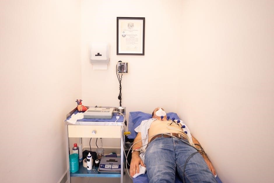
Patient Preparation
Prior to initiating a 12-lead ECG, meticulous patient preparation is paramount for obtaining a clear and interpretable tracing. A comprehensive 12 lead ecg placement pdf will emphasize the importance of explaining the procedure to the patient, alleviating any anxiety and fostering cooperation. Ensure the patient is positioned comfortably, preferably supine, with arms at their sides and legs uncrossed.
Skin preparation is critical; the areas where electrodes will be applied must be clean, dry, and free from lotions, oils, or excessive hair. Shaving may be necessary in areas with abundant hair to ensure optimal electrode contact. Alert the patient to remain still during the recording process, as movement can introduce artifacts and compromise the accuracy of the ECG. Finally, inquire about any implanted devices, such as pacemakers or ICDs, as this will influence electrode placement to avoid interference and ensure patient safety, as detailed in the referenced pdf guide.
Electrode Application – Limb Leads
Correct limb lead placement forms the foundation of an accurate 12-lead ECG. A detailed 12 lead ecg placement pdf will illustrate the standardized color-coding: Red for the right arm (RA), Yellow for the left arm (LA), Green for the left leg (LL), and Black for the right leg (RL) – serving as the ground.
Electrodes should be applied to fleshy areas of the limbs, avoiding bony prominences and muscle contractions. For RA and LA, place electrodes on the lower part of the upper arm, ensuring good skin contact. LL is positioned on the lower calf, while the RL electrode is typically placed on the right upper chest, acting as a ground to minimize interference. Proper adhesion is vital; use appropriate electrode pads and ensure firm contact. Consistent placement, as guided by the pdf, minimizes errors and ensures reliable ECG interpretation.
Electrode Application – Chest Leads
Precise chest (precordial) lead placement, detailed in a comprehensive 12 lead ecg placement pdf, is essential for accurate diagnosis. V1 and V2 are placed in the fourth intercostal space, V1 to the right of the sternum and V2 to the left, directly over the heart. V3 and V4 reside midway between V2 and V4, also in the fifth intercostal space.
V5 is positioned in the anterior axillary line, level with V4, while V6 is placed in the mid-axillary line, at the same intercostal level. Ensuring correct intercostal space identification is crucial – avoid placing electrodes directly on bone or muscle. Good skin contact is paramount; proper electrode adhesion and skin preparation minimize artifact. Referencing a 12 lead ecg placement pdf ensures standardized positioning, leading to reliable and interpretable ECG recordings.

Common Mistakes in ECG Placement
A 12 lead ecg placement pdf highlights frequent errors like incorrect lead positioning, poor skin contact, and improper patient preparation, impacting diagnostic accuracy.
Incorrect Limb Lead Placement
A comprehensive 12 lead ecg placement pdf frequently emphasizes the critical nature of accurate limb lead positioning. Misplacing these electrodes – often due to confusion regarding anatomical landmarks or reversed arm placements – can dramatically alter the ECG tracing, leading to misinterpretations and potentially incorrect diagnoses.
Specifically, swapping the right and left arm leads can invert the P wave and QRS complex in Lead I, mimicking certain arrhythmias. Incorrect placement on the legs can similarly distort the waveform, affecting the interpretation of inferior leads (II, III, aVF). Ensuring proper placement – red on the right arm, yellow on the left arm, green on the left leg, and black on the right leg – is paramount.
The pdf guides often include detailed diagrams and emphasize the importance of verifying placement before initiating the recording, as even slight deviations can introduce significant artifacts and compromise the clinical value of the ECG.
Incorrect Chest Lead Placement
A detailed 12 lead ecg placement pdf consistently highlights the challenges associated with accurate precordial (chest) lead positioning. Unlike limb leads, chest lead placement relies heavily on intercostal space identification and precise anatomical landmarks, making it prone to errors. Common mistakes include placing leads in the wrong intercostal space, or too far medial or lateral from the sternum.
For instance, shifting V1 and V2 too far laterally can diminish R-wave progression, potentially mimicking a posterior myocardial infarction. Incorrect V3 and V4 placement can obscure septal Q waves. A pdf guide will often stress the importance of following the anatomical “landmarks” – the 4th intercostal space for V2, and the 5th intercostal space for V4 – as starting points.
Consistent, standardized placement, as illustrated in the pdf, is vital for reliable ECG interpretation and comparison across different recordings.
Electrode Impedance Issues
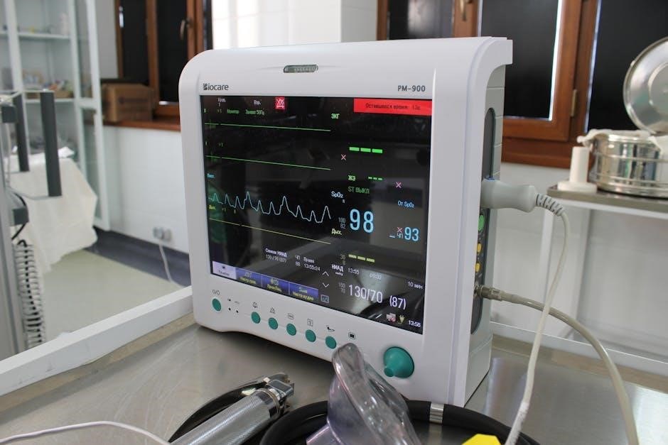
A comprehensive 12 lead ecg placement pdf invariably addresses the critical issue of electrode impedance. High impedance – resistance to the electrical signal – can significantly degrade ECG quality, introducing noise and distorting waveforms. This often stems from poor skin preparation, dried-out electrode gel, or loose electrode contact.
The pdf will typically explain that excessive impedance can lead to a weak or unstable signal, making accurate interpretation difficult. Modern ECG machines often display impedance readings for each lead, alerting technicians to potential problems. Solutions outlined in the pdf include vigorous skin preparation with alcohol wipes, ensuring adequate gel application, and firmly securing electrodes to the skin.
Troubleshooting guides within the pdf emphasize checking connections and replacing electrodes if impedance remains high, ensuring a clear and reliable ECG tracing.

Troubleshooting ECG Signal Quality
A 12 lead ecg placement pdf highlights reducing noise and addressing baseline wander as key to obtaining a clear, interpretable ECG tracing for accurate diagnosis.
Reducing Noise and Artifact
A comprehensive 12 lead ecg placement pdf emphasizes meticulous preparation and technique to minimize interference. Common sources of noise include patient movement, electrical interference from nearby devices, and poor electrode contact. Ensuring the patient is relaxed and still is paramount; explain the procedure clearly to alleviate anxiety which can contribute to muscle tremors.
Proper skin preparation is vital – cleaning the application sites with alcohol to remove oils and dead skin cells improves conductivity. Firmly adhering electrodes with appropriate gel or paste reduces impedance and minimizes artifact. Shielding the ECG machine and patient from external electrical sources, such as IV pumps or monitors, is also crucial. Regularly check electrode connections and replace any that are loose or damaged. Finally, recognizing and documenting artifacts allows for accurate interpretation, avoiding misdiagnosis based on spurious signals.
Addressing Wandering Baseline
A detailed 12 lead ecg placement pdf will highlight the importance of a stable baseline for accurate interpretation. Wandering baseline, often caused by patient movement, respiration, or poor electrode contact, obscures the ECG signal. First, ensure proper patient positioning and minimize movement during recording. Confirm electrodes have secure, low-impedance contact with the skin; re-apply if necessary after thorough skin preparation.
Adjusting the ECG machine’s baseline settings can help, but this is a temporary fix. Identifying and eliminating the source of the interference is key. If respiration is the cause, instructing the patient to briefly hold their breath can stabilize the baseline. Consistent monitoring of electrode quality throughout the recording process is essential. Documenting instances of baseline wander allows for informed interpretation, and recognizing its presence prevents misidentification of true cardiac events.
Resources for 12-Lead ECG Placement (PDFs & Guides)
Numerous online ECG placement guides and downloadable PDF resources are available, offering comprehensive instructions and visual aids for mastering accurate 12-lead ECG technique.
Online ECG Placement Guides
The digital age provides a wealth of accessible online resources dedicated to mastering 12-lead ECG placement. Websites like “The Biomed Guys” offer complete guides encompassing 3-, 5-, and 12-lead electrode positioning, emphasizing accurate technique for precise heart monitoring and diagnosis. These platforms frequently incorporate interactive diagrams and videos, enhancing the learning experience and catering to diverse learning styles.
Many hospital systems and cardiology professional organizations also host online tutorials and placement guides, often tailored to specific equipment or protocols. Searching for “12 lead ecg placement pdf” will yield numerous results, including downloadable guides from reputable sources. These PDFs often include detailed anatomical illustrations, step-by-step instructions, and troubleshooting tips. It’s crucial to verify the source’s credibility and ensure the information aligns with current best practices before relying on any online guide.
Furthermore, several medical education platforms offer comprehensive ECG courses that include dedicated modules on proper lead placement, often accompanied by quizzes and assessments to reinforce learning. Utilizing these resources can significantly improve competency and confidence in performing accurate ECGs.
Downloadable PDF Resources
A search for “12 lead ecg placement pdf” reveals a substantial collection of downloadable resources designed to aid healthcare professionals in mastering this essential skill. These PDFs frequently offer a concentrated and portable format for learning and quick reference, proving invaluable during clinical practice or educational settings.
Many PDFs detail step-by-step instructions accompanied by clear anatomical diagrams illustrating optimal electrode placement for both standard and modified techniques. Some resources focus on common pitfalls and troubleshooting, addressing issues like incorrect lead placement or signal interference. University medical centers and cardiology societies often publish comprehensive guides available for free download.
When selecting a PDF, prioritize resources from recognized medical institutions or professional organizations to ensure accuracy and adherence to current guidelines. Always cross-reference information with established protocols and consider the specific equipment used in your clinical environment. Regularly updating these resources is vital, as best practices evolve.

Advanced Considerations
Special populations, like obese patients, and those with pacemakers/ICDs, require modified 12-lead ECG placement techniques detailed in specialized 12 lead ecg placement pdf guides.
ECG Placement in Specific Populations (e.g., Obese Patients)
Accurate ECG interpretation hinges on proper electrode placement, a challenge amplified in specific patient populations. Obese patients, for instance, often present difficulties due to subcutaneous fat potentially distorting electrical signals and shifting electrode positions. Standard placement may necessitate adjustments; precordial leads might require positioning in intercostal spaces lower than typically recommended to ensure contact with the heart.
Furthermore, utilizing larger electrodes can improve skin contact and signal quality. A comprehensive 12 lead ecg placement pdf guide will illustrate these modified techniques. Patients with unusual body habitus or anatomical variations also demand individualized approaches. Careful consideration of chest wall curvature and muscle mass is vital. Always prioritize maximizing signal clarity while adhering to established guidelines, consulting specialized resources when encountering complex cases. Proper skin preparation is also crucial for optimal adhesion and signal acquisition in these patients.
ECG Placement and Pacemakers/ICDs
Patients with implanted pacemakers or implantable cardioverter-defibrillators (ICDs) require modified ECG placement protocols. The device itself can create artifact, obscuring the underlying electrical activity. Chest leads should be positioned at least 2-3 inches away from the device generator to minimize interference. A detailed 12 lead ecg placement pdf resource will demonstrate optimal lead positioning relative to these devices;
Avoid placing electrodes directly over the device or its leads. The presence of a pacemaker or ICD can alter the typical ECG waveform, necessitating careful interpretation. Understanding the device’s function and lead configuration is crucial. It’s important to document the device’s presence and location on the ECG report. Specialized techniques may be needed to differentiate device-related signals from intrinsic cardiac activity, ensuring accurate diagnosis and treatment planning. Always prioritize patient safety and consult relevant guidelines.

Future Trends in ECG Technology
The future of ECG technology is rapidly evolving, moving beyond traditional 12-lead systems. Wireless and wearable ECG monitors are gaining prominence, offering continuous monitoring and remote data transmission. Artificial intelligence (AI) and machine learning algorithms are being integrated to enhance ECG interpretation, improving diagnostic accuracy and efficiency. These advancements will necessitate updated training materials, and a comprehensive 12 lead ecg placement pdf will need to reflect these changes.
Expect to see more sophisticated algorithms for automated ECG analysis and real-time arrhythmia detection. Integration with telehealth platforms will enable remote patient monitoring and timely intervention. Research is focused on developing more user-friendly and accessible ECG devices. The focus will shift towards preventative cardiology, utilizing ECG data for risk stratification and personalized medicine. Staying current with these trends is vital for healthcare professionals.